|
|
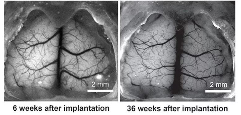
Researchers at University of Minnesota have created See-Shell, a transparent skull for studying neural activity in mice. The use of such monitoring tools to measure neural activity in real time is providing massive insight into the treatment of concussions, Alzheimer’s and other neural disorders. The device is as simple as a replacement for the regular skull, allowing researchers to image the organ periodically. In due time, the team also hopes it will aid in improving treatments based on their findings. See-Shell allows researchers to monitor the activity within mice to get a clear picture of how activity in one portion of the brain is tied to other parts of the organ.
“These are studies we couldn’t do in humans, but they are extremely important in our understanding of how the brain works so we can improve treatments for people who experience brain injuries or diseases,” says Timothy J. Ebner. Each See-Shell skull enables the team to fit “penetrating neural probes to perturb or record neural activity simultaneously with whole cortex imaging” according to the study. More specifically, the skulls provide roughly 300 days of optical monitoring access to 45 mm2 of dorsal tissue within the mice. Video research using the device detailed changes in brightness in response to heightened neural activity. Subtle flashes appear when the whole brain fires up at once.
The researchers detail their findings in a study they published in Nature, ‘Cortex-wide neural interfacing via transparent polymer skulls‘. They constructed the See-Shells by imaging the real skulls of the mice and recreating them via a common desktop 3D printer. The frame of the skull consists of printed polymethylmethacrylate (PMMA) with a thin, flexible and transparent PET film. PET and PMMA are ideal for this sort of experiment as they are sufficiently transparent and easy to print. “This will give us new information about how the human brain works. This technology allows us to see most of the cortex in action with unprecedented control and precision while stimulating certain parts of the brain,” Suhasa Kodandaramaiah, a co-author of the study, said
While mouse brains are certainly different, they provide some useful similarities to human neurology. They hope that the answers within mice will be sufficiently applicable to the medical field. The researchers are currently using the See-Shells to study Parkinson’s, Alzheimer’s and a range of other diseases. They also looked into how mild concussions spread throughout the brain and how it recovers. Featured image and video courtesy of University of Minnesota.
